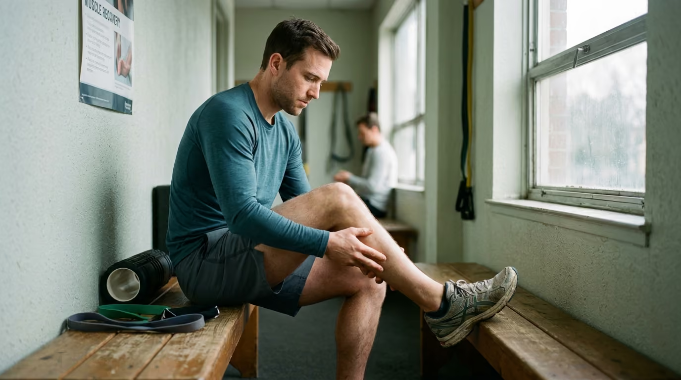

Stiff morning joints and persistent tendon discomfort require progressive mechanical loading protocols, targeted regional exercises.





You have likely searched for why your Achilles tendon aches every morning, why your elbow flares during heavy pulling, or why your shoulder throbs after overhead work. You probably found conflicting advice telling you to rest completely, apply ice, stretch aggressively, or buy specialized supplements. This guide provides a definitive, research led blueprint for building durable connective tissue based on mechanical loading, tissue capacity, and biological adaptation.
Connective tissue does not behave like muscle tissue. Tendons adapt along a different timeline, respond to specific mechanical strains, and require deliberate loading to remodel effectively. Understanding these mechanisms allows you to build long term resilience without wasting time on passive treatments that fail to build capacity.
A tendon is specialized connective tissue designed to transmit muscular force to bone while storing and releasing elastic energy. Mature tendon tissue consists predominantly of dense Type I collagen fibers aligned along the primary line of tension. Surrounding these fibers is an extracellular matrix composed of proteoglycans, glycoproteins, water, and specialized cells known as tenocytes. Tenocytes sense mechanical forces through mechanotransduction and convert physical strain into cellular remodeling signals.
When a tendon experiences mechanical loading, tenocytes initiate both collagen synthesis and collagen degradation. Research shows that acute exercise elevates the fractional synthetic rate of tendon collagen from roughly 0.05 percent per hour to nearly 0.10 percent per hour within 24 hours. A measurable increase in synthesis can appear as early as six hours following intense loading. However, collagen degradation also rises acutely following exercise. A net gain in collagen structure only occurs when sufficient recovery allows synthesis to outpace degradation.
Chronic resistance training changes both the material properties and the architecture of the tendon over several months. A landmark systematic review and meta-analysis led by Sebastian Bohm and colleagues examined human tendon adaptation to mechanical loading. The researchers found that mechanical loading produces moderate to large improvements in tendon stiffness and Young's modulus, alongside smaller changes in cross sectional area. Across the reviewed literature, standardized mean differences reached 0.70 for tendon stiffness, 0.69 for Young's modulus, and 0.24 for cross sectional area.
These findings show that a resilient tendon is not merely a thicker tendon. Structural hypertrophy of connective tissue occurs slowly and modestly. The primary driver of increased tendon stiffness is an improvement in the material quality of the extracellular matrix itself. High magnitude loading increases cross linking between collagen molecules, packs fibrils more densely, and alters the fluid dynamics of the tissue.
To build durable physical capacity, your training must target the mechanical properties of the tendon rather than chasing muscle fatigue alone. Readers looking to build robust physical structures should integrate these principles into their wider physical performance and strength protocols.
One of the most common pitfalls in connective tissue management is confusing symptom relief with structural remodeling. Tendon pain, structural appearance on imaging, mechanical stiffness, and functional capacity operate on distinct physiological timelines.
In clinical studies of midportion Achilles tendinopathy, patients frequently experience meaningful reductions in pain and improvements in daily function within two to four weeks of beginning a loading program. However, measurable muscular hypertrophy generally requires at least four weeks of structured resistance. Meaningful structural remodeling of the tendon matrix often takes twelve weeks to several months of consistent mechanical strain.
If you return to high-impact sprinting, jumping, or heavy lifting the moment your pain subsides, you place high tensile forces on an unadapted matrix. The surrounding muscle may be capable of producing force, but the tendon has not yet developed the modulus or strain tolerance to handle repeated cycles.
Structural abnormalities on medical imaging also correlate poorly with actual performance and pain. Up to 25 percent of completely asymptomatic athletes display structural abnormalities, hypoechoic areas, or thickening on ultrasound and MRI scans. Conversely, an individual can experience debilitating localized tendon pain with minimal visible changes on standard imaging.
Current clinical practice guidelines recommend against routine imaging for standard tendon complaints. Imaging should be reserved for diagnostic uncertainty, failure to respond to progressive conservative loading, or suspicion of an acute structural rupture.
Tendons respond specifically to the magnitude, rate, and duration of the strain applied to them. Research indicates that high magnitude loading, generally defined as resistance training performed at or above 70 to 85 percent of maximum voluntary contraction, produces superior tendon adaptations compared to low load resistance training.
Low load training with high repetitions may build muscular endurance, but it generates insufficient strain to trigger significant tenocyte mechanotransduction and collagen synthesis.
A comprehensive tendon resilience strategy moves deliberately through three distinct loading regimes.
Isometric contractions produce muscular tension without changing joint angles or tendon length. This loading style provides a stable entry point when a tendon is reactive, painful, or intolerant of dynamic movement.
Isometric contractions allow you to apply high mechanical force to the tendon while avoiding peak compressive forces that occur at the extremes of joint ranges.
In clinical frameworks, sustained isometric holds lasting 30 to 45 seconds, repeated four to five times per session at roughly 70 percent of maximum effort, can provide a predictable loading stimulus.
Isometrics are an effective tool for maintaining force production capacity during irritable phases. However, they should not become a permanent substitute for dynamic strength and energy storage work.
Heavy slow resistance involves controlled concentric and eccentric contractions using external resistance. Repetitions are executed with a deliberate cadence, typically taking three seconds to lift and three seconds to lower.
This tempo eliminates momentum, ensures continuous tension across the entire range of motion, and maximizes the time under tension for the connective tissue.
A systematic review published in the Journal of Orthopaedic and Sports Physical Therapy confirmed that heavy slow resistance produces clinical and structural outcomes comparable or superior to traditional isolated eccentric training. Heavy slow training improves tendon stiffness, increases tensile tolerance, and builds the muscular strength required to shield the tendon from excessive strain rates.
Tendons function as biological springs during locomotion, sprinting, jumping, and rapid changes of direction. During the stretch shortening cycle, the muscle contracts quasi-isometrically while the tendon rapidly elongates and recoils to propel the body.
Slow resistance training builds the material strength of the tendon, but it does not prepare the tissue for the high strain rates experienced during explosive movement.
Once adequate baseline strength and stiffness are established, your program must introduce progressive plyometric and high-rate activities. This includes skipping, linear hopping, multi-directional bounding, and sport-specific acceleration drills. Progressing into energy storage work ensures that the muscle-tendon complex can absorb and release elastic energy efficiently.
Managing connective tissue requires balancing mechanical stimulus with adequate biological recovery. Complete rest is counterproductive for chronic tendon issues. Immobilization or prolonged unweighting causes rapid catabolism of the tendon matrix, decreases collagen synthesis, and reduces mechanical stiffness. The goal is relative load modification: keeping the tendon active within a tolerable envelope while systematically increasing capacity.
Pain during training is acceptable if it remains within controlled thresholds. In research frameworks for Achilles and patellar tendinopathy, pain levels up to 4 or 5 on a 10-point scale during exercise are considered acceptable, provided that symptoms return to baseline within 24 hours.
The next-morning assessment provides your most reliable data point. Tendons typically react to excessive volume or excessive strain rates several hours after the session. If you wake up with increased tendon stiffness, localized tenderness, or pain during your initial morning steps, your previous day's training exceeded your current tissue capacity.
When a negative reaction occurs, do not cease training entirely. Simply reduce one training variable in the subsequent session. You can decrease the movement speed, reduce the total number of sets, or limit the range of motion while maintaining the resistance. Managing this biological response is closely tied to systemic recovery strategies that support tissue repair.
Different anatomical regions impose distinct mechanical constraints on connective tissue. A durable tendon program must account for local joint biomechanics, compressive forces, and the primary demands of the target movement.
The Achilles tendon experiences some of the highest mechanical loads in the human body, enduring forces up to eight times body weight during running and jumping.
Achilles issues generally fall into two anatomical categories: midportion tendinopathy, located two to six centimeters above the heel bone, and insertional tendinopathy, located directly at the calcaneus attachment.
Insertional tendinopathy is sensitive to compressive loads. Performing calf raises with the heels dropping below a step can compress the tendon against the calcaneus, aggravating symptoms. Insertional protocols should initially be performed on flat ground. Midportion issues, by contrast, benefit from moving through full dorsiflexion and plantarflexion as tissue capacity improves.
Lateral elbow pain, often termed tennis elbow, involves the common extensor tendon origin, particularly the extensor carpi radialis brevis. This tissue is heavily stressed by repetitive gripping, typing, manual labor, and heavy pulling exercises like deadlifts and rows.
Clinical guidelines from the American Physical Therapy Association emphasize that lateral elbow rehabilitation should not be restricted to isolated wrist movements. The program must strengthen the entire kinetic chain, including the rotator cuff and scapular stabilizers, while applying direct load to the wrist extensors.
The rotator cuff consists of the supraspinatus, infraspinatus, teres minor, and subscapularis. These tendons dynamically stabilize the humeral head within the glenoid fossa during pressing, pulling, and overhead movements.
Rotator cuff tendinopathy frequently develops from rapid spikes in pressing volume, prolonged overhead work, or insufficient strength in external rotation relative to internal rotation.
A 2025 clinical practice guideline confirmed that progressive resisted exercise significantly reduces pain and restores shoulder function. Effective shoulder rehabilitation must expose the rotator cuff to resistance across multiple planes while reinforcing the posterior upper back.
Integrating these exercises into your broader training split establishes a durable foundation that supports healthy aging and physical longevity.
High-performing professionals often struggle to maintain rigid rehabilitation or strength programs while navigating demanding work schedules, frequent flights, and extended hotel stays. When you lack access to specialized gym equipment, you can still apply meaningful mechanical strain to your connective tissue using body weight, long isometric holds, and portable resistance bands.
I spent a week at a popular health optimization conference and left completely exhausted by the complexity. Everyone was pushing a new supplement protocol, a complicated gadget, or a rigid daily routine. It struck me that true high performers do not have time to make health a full time job. They need maximum return on minimum viable effort. That observation became the filter for every piece of research we publish across our curated executive performance resources.
Tendon tissue does not know the difference between a high-end plate-loaded machine and a rigorous bodyweight isometric setup. The tenocytes only register the mechanical tension and cellular strain placed across the extracellular matrix.
During periods of intense cognitive stress and heavy travel, systemic recovery resources are compromised. High stress elevates systemic cortisol and inflammatory cytokines, which can slow down local matrix remodeling.
When your travel schedule intensifies, reduce the total weekly volume of tendon loading by 30 to 50 percent while preserving the mechanical intensity of your exercises. This maintains connective tissue stiffness without overloading your nervous system, a principle explored in our guides on managing sustained physiological stress.
Nutritional interventions for tendon resilience are often marketed with aggressive claims that outpace the underlying scientific data. It is critical to separate established nutritional support from speculative supplementation.
Tendon remodeling depends fundamentally on adequate energy availability and total daily protein intake. Tendon synthesis requires a continuous supply of amino acids, particularly glycine, proline, and hydroxyproline. If an individual is in a chronic energy deficit or consumes insufficient protein, collagen synthesis rates decline regardless of the training stimulus.
Several randomized trials have examined whether taking hydrolyzed collagen or gelatin combined with Vitamin C prior to exercise accelerates tendon adaptation.
A study evaluating female athletes undergoing high-intensity resistance training reported a 15.4 percent increase in patellar tendon stiffness in the collagen supplemented group compared to a 4.6 percent increase in the control group.
The biological rationale is that consuming specific collagen peptides roughly 45 to 60 minutes prior to loading maximizes peak plasma concentrations of glycine and proline precisely when blood flow to the tendon is elevated by exercise.
However, supplementation without mechanical loading produces zero measurable changes in tendon stiffness or strength. Collagen supplements are not a stand-alone therapy. They function at best as a modest nutritional amplifier for an appropriately structured mechanical loading protocol.
While progressive mechanical loading is the gold standard for connective tissue resilience, several physiological boundaries and diagnostic caveats must be acknowledged.
First, mechanical loading protocols exhibit wide individual variability. Systematic reviews show that while the average response to resistance training is an increase in tendon stiffness, individual responses range from substantial improvements to minimal measurable change over short intervention periods. Genetic differences in collagen structure, vascularity, hormonal status, and lifetime training exposure all influence adaptation rates.
Second, systemic metabolic conditions significantly alter tendon biology. Type 2 diabetes, chronic insulin resistance, and dyslipidemia lead to the accumulation of advanced glycation end products within the extracellular matrix. These cross links make the tendon brittle and alter normal cellular signaling, increasing the risk of tendinopathy and tendon rupture.
Third, specific pharmaceutical agents carry well documented toxicities for connective tissue. Fluoroquinolone antibiotics (such as ciprofloxacin and levofloxacin) inhibit tenocyte metabolism and significantly increase the risk of acute tendon rupture, even months after completing the medication course. Similarly, local corticosteroid injections around weight-bearing tendons can provide short-term pain relief at the expense of long-term cellular viability, collagen necrosis, and heightened rupture risk.
Finally, sudden sharp pain accompanied by an audible pop, visible deformity, or immediate loss of motor function indicates an acute full thickness tear or structural rupture. These cases require immediate orthopedic evaluation rather than conservative self-directed loading.
To maintain long term structural durability, tendon health must be approached through a holistic performance framework. Connective tissue does not function in isolation; it operates as an integral part of your musculoskeletal and metabolic architecture.
Building durable connective tissue requires balancing targeted tissue loading with full body strength, mobility, and metabolic health. As detailed in our comprehensive long term musculoskeletal healthspan analysis, maintaining tissue capacity across decades ensures sustained mobility and performance.
By systematically addressing each tier of this matrix, you create an internal environment that supports continuous tissue remodeling. This structured approach allows you to pursue aggressive physical goals while minimizing your vulnerability to chronic connective tissue degradation.
To integrate these principles into your current weekly schedule, apply this practical checklist starting today.
Stay connected for research and practical guidance on executive performance, energy, focus, sleep, recovery and longevity. Ideas built for people who want to stay sharp, capable and effective for the long run.


Build habits and systems that support clear thinking, steady energy and long term capacity throughout a demanding career.
explore the Blog